Brittany: Difference between revisions
imported>Psych204B No edit summary |
imported>Psych204B |
||
| (11 intermediate revisions by the same user not shown) | |||
| Line 29: | Line 29: | ||
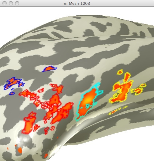
[[File:Face Selective ROIs Final (Localizer).jpg | Figure 1]] | [[File:Face Selective ROIs Final (Localizer).jpg | Figure 1]] | ||
FIGURE 1 | FIGURE 1 | ||
ROIs are named as follows: Face Selective 1- Blue | |||
Face Selective 2- Red | |||
Face Selective 3- Cyan | |||
Face Selective 4- Yellow | |||
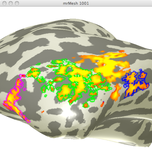
Using the localizer data for limb selective regions from the same experiment I selected 3 limb selective ROIs (See figure 2). I chose these 3 ROIs in order to obtain data from one ROI in 3 differing anatomical regions. | Using the localizer data for limb selective regions from the same experiment I selected 3 limb selective ROIs (See figure 2). I chose these 3 ROIs in order to obtain data from one ROI in 3 differing anatomical regions. | ||
| Line 36: | Line 39: | ||
[[File:Limb Selective ROIs Final.png | FIGURE 2]] | [[File:Limb Selective ROIs Final.png | FIGURE 2]] | ||
FIGURE 2 | FIGURE 2 | ||
ROIs are named as follows: Limb Selective 1- Blue | |||
Limb Selective 2- Green | |||
Limb Selective 3- Magenta | |||
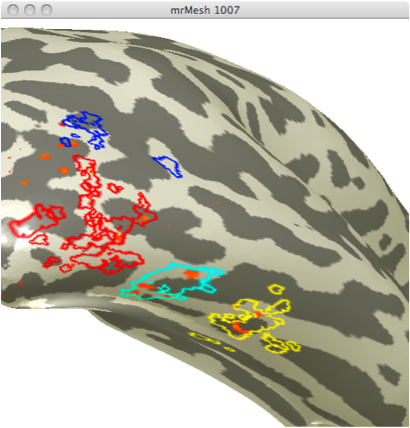
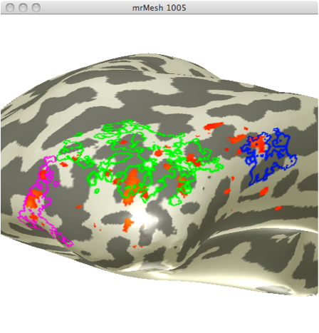
These ROIs were then uploaded onto their respective adaptation maps obtained from the first experiment. Face Selective adaptation map(figure 3) and Limb selective adaptation map (figure 4). | These ROIs were then uploaded onto their respective adaptation maps obtained from the first experiment. Face Selective adaptation map(figure 3) and Limb selective adaptation map (figure 4). | ||
| Line 43: | Line 49: | ||
FIGURE 3 FIGURE 4 | FIGURE 3 FIGURE 4 | ||
The time course for each ROI was then extracted from the adaptation data in order to compare the adaptation between categories, ROIs, and anatomical regions. | |||
= Results = | |||
==Face Selective Time Courses== | |||
[[File:FS1 Timecourse2.png | Figure 5]] Face Selective 1 (Blue) Time course | |||
[[File:FS2Timecourse.png | Figure 6]] Face Selective 2 (Red) Time course | |||
[[File:FS3Timecourse.png | Figure 7]] Face Selective 3 (Cyan) Time course | |||
[[File:FS4Timecourse.png | Figure 8]] Face Selective 4 (Yellow) Time course | |||
As seen from these four time courses, all of ROIs based on faced selective localizer data remains category selective within the adaptation parameter map. Face selective ROI 1 does not show adaptation in response to faces, and furthermore shows a higher response to repeated limb stimuli than non repeated limb stimuli (both responses still less than those for faces). Face selective ROIs 1,2, and 3 show increasing levels of adaptation to all stimuli, excluding houses. | |||
== Limb Selective Time Courses == | |||
[[File:LS1Timecourse.png | Figure 9]] Limb Selective 1 (Blue) Time course | |||
[[File:LS2Timecourse.png | Figure 10]] Limb Selective 2 (Green) Time course | |||
[[File:LS3Timecourse.png | Figure 11]] Limb Selective 3 (Magenta) Time course | |||
Similarly to face selective regions, all limb selective ROIs maintain a significant preference for images of limbs. However, unlike the anterior to posterior decrease in fMRI adaptation seen in face selective regions, the limb selective regions do not show this pattern. In fact, there is a slight increase in adaptation strength from the anterior to posterior limb selective ROIs. | |||
= Conclusions = | = Conclusions = | ||
My results show that fMRI adaptation is significant for both preferred and non-preferred categories, excepting face selective ROI 1. According to the original study from which this data was obtained, this result is expected across "the majority of subjects"[1]. However, the original study also concluded that the largest adaptation should occur within the preferred category which was a result only observed in face selective region 4. | |||
My results also show a decreasing fMRI adaptation ratio (Repeat/Non-repeat) from posterior to anterior regions within the face selective category, indicating a stronger adaptation response in the more anterior regions. This result is expected based on the original study, however, this was also expected but not observed for limb selective regions. This could be due to the fact that I only analyzed one hemisphere of one participant. Additionally, the selection of only three limb ROIs and my selection of ROI boundaries could have greatly influenced this result. As a next step I would go back and analyze additional ROIs within the limb selective regions in order to gain a better understanding of the fMRI adaptation patterns within that category's data. | |||
= | = References = | ||
1) Sayres & Grill-Spector, J. Neurophysiology, 2006 | |||
2) Weiner K., Sayres R., Vinberg J., Grill-Spector K. fMRI Adaptation and Category Selectivity in Human Ventral Temporal Cortex: Regional Differences Across Time Scales. J. Neurophysiology, 2010 | |||
3) Weiner K., Grill-Spector K.. Sparsely distributed organization of face and limb activations in human ventral temporal cortex. Neuroimage, 2010 | |||
Latest revision as of 16:12, 15 March 2012
The Relationship between fMRI adaptation and category selectivity
This project investigates the link between adaptation and areas of the cortex linked to face recognition and limb recognition using an event related design.
Background
fMRI Adaptation is a phenomena that takes place in higher order cortical areas in which the BOLD response signal decreases in response to repeated identical visual stimuli. Adaptation has been shown to occur in higher order visual areas but not early visual areas such as V1 [1]. This study investigates the relationship between object selectivity and fMRI adapation. More specifically, it will examine face selective areas as well as body part selective areas with regards to adaptation. Furthermore, it will look at the differences between adaptation within the ROIs of those two specific categories as well as the similarities in adaptation across various anatomical brain areas.
Methods
Pre-processed data was used from the lab of Kalanit Grill-Spector from the parts of an experiment outlined below [2].
Subjects
The data from the right hemisphere of one subject, a male in his late 20s, was analyzed.
MR acquisition
The subject participated in two experiments, an event related design followed by a block design localizer scan. The first, event related design, consisted of 8 runs of 156 trials lasting for 2 seconds each. Each trial consisted of the presentation of an image of either a face, limb, car, or house for 1000ms followed by a 1000ms blank. Within each run, only two of the images were repeated six times, the rest not repeated at all. None of the images were repeated across scans.
The second experiment was a block design used to identify category selective areas within the ventral stream. Blocks were 12 seconds long with a 750ms stimulus presentation period followed by a 250 ms blank period. Each run consisted of 32 blocks, 4 blocks for each category (faces, limbs, flowers, cars, guitars, houses, and scrambled) as well as four blank blocks.
12 slices were acquired at a resolution of 1.5 x 1.5 x 3mm per voxel and a TR of 1000 ms.
MR Analysis
As mentioned above, the data I analyzed was pre-processed. That high resolution MR data was was then analyzed using mrVista software tools.
ROIs
Using the localizer data from the second experiment, I created ROIs specific for areas selective for faces and areas selective for limbs. I chose the 4 different face selective areas seen in figure 1.
                    ROIs are named as follows: Face Selective 1- Blue
                                               Face Selective 2- Red
                                               Face Selective 3- Cyan
                                               Face Selective 4- Yellow
Using the localizer data for limb selective regions from the same experiment I selected 3 limb selective ROIs (See figure 2). I chose these 3 ROIs in order to obtain data from one ROI in 3 differing anatomical regions.
                    ROIs are named as follows: Limb Selective 1- Blue
                                               Limb Selective 2- Green
                                               Limb Selective 3- Magenta
These ROIs were then uploaded onto their respective adaptation maps obtained from the first experiment. Face Selective adaptation map(figure 3) and Limb selective adaptation map (figure 4).
FIGURE 3 FIGURE 4
The time course for each ROI was then extracted from the adaptation data in order to compare the adaptation between categories, ROIs, and anatomical regions.
Results
Face Selective Time Courses
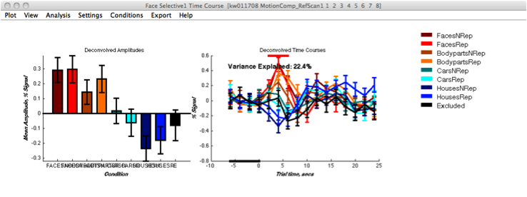 Face Selective 1 (Blue) Time course
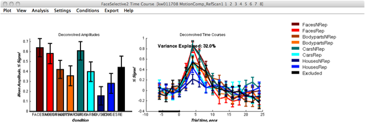 Face Selective 2 (Red) Time course
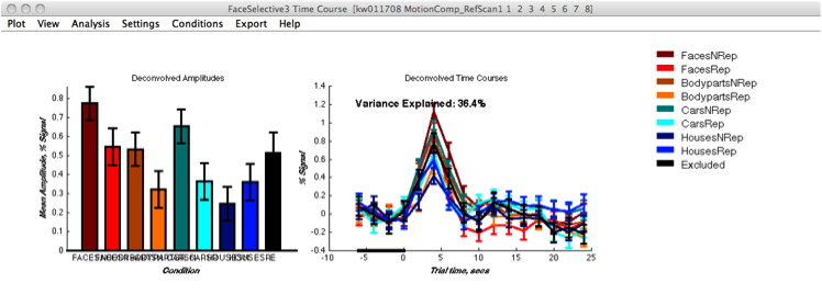 Face Selective 3 (Cyan) Time course
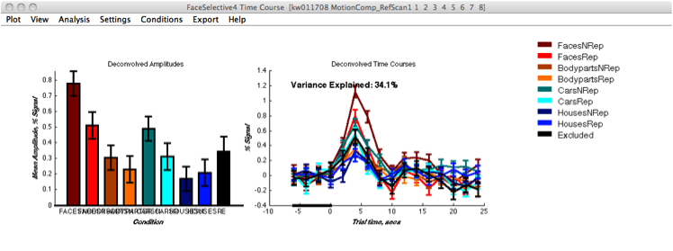 Face Selective 4 (Yellow) Time course
As seen from these four time courses, all of ROIs based on faced selective localizer data remains category selective within the adaptation parameter map. Face selective ROI 1 does not show adaptation in response to faces, and furthermore shows a higher response to repeated limb stimuli than non repeated limb stimuli (both responses still less than those for faces). Face selective ROIs 1,2, and 3 show increasing levels of adaptation to all stimuli, excluding houses.
Limb Selective Time Courses
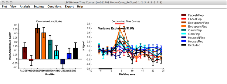 Limb Selective 1 (Blue) Time course
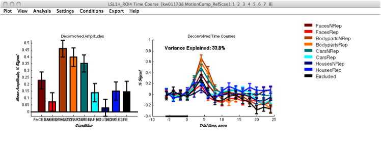 Limb Selective 2 (Green) Time course
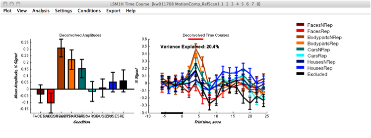 Limb Selective 3 (Magenta) Time course
Similarly to face selective regions, all limb selective ROIs maintain a significant preference for images of limbs. However, unlike the anterior to posterior decrease in fMRI adaptation seen in face selective regions, the limb selective regions do not show this pattern. In fact, there is a slight increase in adaptation strength from the anterior to posterior limb selective ROIs.
Conclusions
My results show that fMRI adaptation is significant for both preferred and non-preferred categories, excepting face selective ROI 1. According to the original study from which this data was obtained, this result is expected across "the majority of subjects"[1]. However, the original study also concluded that the largest adaptation should occur within the preferred category which was a result only observed in face selective region 4.
My results also show a decreasing fMRI adaptation ratio (Repeat/Non-repeat) from posterior to anterior regions within the face selective category, indicating a stronger adaptation response in the more anterior regions. This result is expected based on the original study, however, this was also expected but not observed for limb selective regions. This could be due to the fact that I only analyzed one hemisphere of one participant. Additionally, the selection of only three limb ROIs and my selection of ROI boundaries could have greatly influenced this result. As a next step I would go back and analyze additional ROIs within the limb selective regions in order to gain a better understanding of the fMRI adaptation patterns within that category's data.
References
1) Sayres & Grill-Spector, J. Neurophysiology, 2006
2) Weiner K., Sayres R., Vinberg J., Grill-Spector K. fMRI Adaptation and Category Selectivity in Human Ventral Temporal Cortex: Regional Differences Across Time Scales. J. Neurophysiology, 2010
3) Weiner K., Grill-Spector K.. Sparsely distributed organization of face and limb activations in human ventral temporal cortex. Neuroimage, 2010
