Feng: Difference between revisions
imported>Student221 |
imported>Student221 |
||
| Line 38: | Line 38: | ||
With the OralEyeCamera.exe command line tool, we were able to capture sequences of frames with different lighting/exposure at a frame rate of about 2 fps. The frame rate is mainly limited by the overhead of file I/O, establishing socket communication and parameter setting for each frame. However, for a consecutive capture of 6 frames, the subject only needs to keep the mouth open for ~3 seconds, which is still feasible in practice. | With the OralEyeCamera.exe command line tool, we were able to capture sequences of frames with different lighting/exposure at a frame rate of about 2 fps. The frame rate is mainly limited by the overhead of file I/O, establishing socket communication and parameter setting for each frame. However, for a consecutive capture of 6 frames, the subject only needs to keep the mouth open for ~3 seconds, which is still feasible in practice. | ||
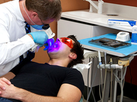
[[File:Subject001.png|thumb|upright=1.5|left|alt=Alt text|Figure 5: Subject 001 with white and blue illumination.]] | [[File:Subject001.png|thumb|upright=1.5|left|alt=Alt text|Figure 5: Subject 001 with white and blue illumination.]] | ||
A pair of images of subject 003 are shown in Figure 4. | |||
== Conclusions == | == Conclusions == | ||
Revision as of 05:01, 14 December 2019
Introduction
Cancer screening is a procedure that aims to detect cancer before any signs or symptoms of cancer arise; it is potentially useful for medical diagnosis because early detection of cancer significantly increases the chances for successful treatment [1]. For the case of oral cancer, screening is particularly important as survival does correlate with stage, making early diagnosis and treatment optimal for this disease [2]. Unfortunately, the diagnosis constantly rely on physical examination followed by biopsy confirmation, which could result in delay in diagnosis [3]. As such, more and more research are being conducted to develop easy-to-use, cost effective oral cancer screening devices to encourage and improve the screening process.
Currently, there is a number of devices available on the market for oral cancer screening purpose. Some examples are Velscope®, ViziLite® and Identafi®. This project is based on the newly developed "OralEye" system from Fengyun Vision Technology, and aims to improve the overall usability, accuracy and efficiency of the system for oral screening.
Background
This section provides an overview of existing oral cancer screening devices as well as areas of improvements proposed by researchers, which will be serving as guidance for development of the new screening system based on OralEye camera.
ViziLite is a disposable capsule like device that relies on chemiluminescence in 430-580 nm. Although researchers found its potential utility in identifying occult epithelial abnormalities, ViziLite suffers from high false positive and negative levels, and thus limiting its clinical application [4].

VELscope® is a hand held device that emits 400–460 nm wavelength light to excite auto-fluorescence from fluorophores in the mouth. Some researchers suggest that 405 nm wavelength light was able to discriminate neoplastic and non-neoplastic tissue with high sensitivity and specificity [5]. However, there has also been a number of criticisms on VELscope® for the limited capacity to extend its use in general dentistry. Researchers are still trying to improve its specificity to allow wider clinical use [4].
Identafi® is a probe like device designed for multi-spectral screening of oral disease. It emits three types of light: white, violet (405 nm) and green-amber (545 nm) light. The white light provides classical visual inspection and the violet light is for similar purpose as in VELscope®. The green-amber light, through reflection spectroscopy, is used to excites haemoglobin molecules in the blood, in order to visualise the vasculature [6]. Researchers found that the use of Itentafi provides more data that conventional oral exam. However, it requires high level of clinical training in oral pathology to interpret the results, and thus limiting its usage in general practice [7].
According to ref [4], a main limiting factor for universal usage of these devices is no demonstrated superiority compared to conventional oral exam. However, the authors acknowledge the possibility that these devices can be enhanced with "new approaches used to analyze optical imaging data". As such, this project aims to explore new methods towards easy-to-use oral florescence imaging as well as accurate data interpretation to address the needs in oral cancer screening.
Methods

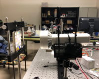
The OralEye camera has a form factor similar to that of a torch or flashlight, and is mounted on an optical post to take images of subjects or calibration targets. The camera is connected to a PC using a USB cable. The communication between the camera and PC is based on the Ethernet-over-USB protocol RNDIS. A software development kit (SDK) provided by Fengyun Vision is used to develop custom camera interfacing software. A basic illustration of the communication is shown in Figure 3.

The command line camera control software OralEyeCamera.exe is developed in Visual C++ with Visual Studio 2017. Most of the communication between the camera and PC is based on the TCP protocol. As such, the Winsock library is heavily used. The executable can take four types of input parameters to carry out different tasks. The syntax for using the command line tool is shown below:
OralEyeCamera.exe printParam
OralEyeCamera.exe lightBlue|lightWhite|lightOff
OralEyeCamera.exe blueShoot|whiteShoot|singleShoot [expTime]
OralEyeCamera.exe task.json
The JSON file is used to specify a sequence of captures with different light/exposure settings. The JSON file format is defined in the separate MATLAB software "oraleye".
Results

With the OralEyeCamera.exe command line tool, we were able to capture sequences of frames with different lighting/exposure at a frame rate of about 2 fps. The frame rate is mainly limited by the overhead of file I/O, establishing socket communication and parameter setting for each frame. However, for a consecutive capture of 6 frames, the subject only needs to keep the mouth open for ~3 seconds, which is still feasible in practice.

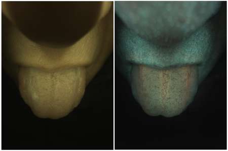
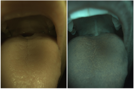
A pair of images of subject 003 are shown in Figure 4.
Conclusions
Appendix
You can write math equations as follows:
You can include images as follows (you will need to upload the image first using the toolbox on the left bar, using the "Upload file" link).
References
[1] https://www.who.int/cancer/detection/en/
[2] Ries LAG, Kosary CL, Hankey BF, et al. SEER Cancer Statistics Review, 1973-1995. Bethesda, MD, NCI.2. American Cancer Society, Facts and Figures, 2000.
[3] https://oralcancerfoundation.org/discovery-diagnosis/cancer-screening-protocols/
[4] Mascitti, M., et al. "An Overview on Current Non-invasive Diagnostic Devices in Oral Oncology" Front. Physiol. 9, 1510 (2018)
[5] Roblyer, D., et al. "Objective detection and delineation of oral neoplasia using autofluorescence imaging", Cancer Prev. Res. 2, 423–431 (2009)
[6] Messadi, D. V., et al. "The clinical effectiveness of reflectance optical spectroscopy for the in vivo diagnosis of oral lesions", Int. J. Oral Sci. 6, 162–167 (2014)
[7] Lalla, Y., et al. "Assessment of oral mucosal lesions with autofluorescence imaging and reflectance spectroscopy", J. Am. Dent. Assoc. 147, 650–660.

