Neuroimaging
Back to Psych 221 Projects 2013
Neuroimaging
Introduction
With the opening of Stanford's Center for Cognitive and Neurobiological Imaging (CNI), we now have access to a large number of MR scans of the human brain. As they are also closely connected to the MR hardware and image processing algorithms, the CNI, being a shared facility that is dedicated to research and teaching, provides resources for researchers and students used in cognitive and neurobiological sciences. The MR images that were used for this project were obtained courtesy of the CNI.
Motivation
Brain vs Squash
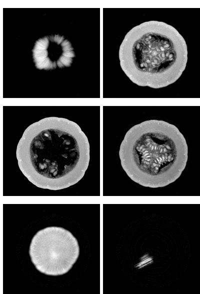
In the field of medicine, the medical students start out to take effective MR scans by first practicing on inanimate objects such as a squash or pumpkins. However, it so happens, that these MR images used for practice, often get stored into the real database of actual human MR images. This proves to be a major problem and hence must be rectified without the need to manually go through all the scans. Sometimes the scans are labelled incorrectly, thus causing the contents to be totally different from the label and hence be a misleading piece of data. That was the main motivation behind the project; to be able to separate the scans of the squash from that of the head so that there wouldn't be trivial errors while keeping and maintaining records at hospitals.


Brain vs Brain
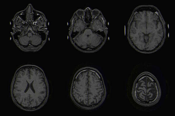
Efficiently and accurately matching the MRI of brain in database is very important for doctors to find the similar cases. However the definition of similarity of cases may vary according to different doctors’ experience. A matching algorithm for brain MRI is presented in this paper, with the semantic label consisting of the number of regions, the size, the location and the shape feature of each region of the MRI of brain. Future more, to adapt doctors’ preferences, the relevance feedback is taking into consideration to update the weight of each feature and to save the doctors’ preferences. The testing results show that the algorithm gives a good performance.
Project Aim
Thus the project was divided into two halves.
a) Brain vs Squash detection
b) Brain vs Brain detection
Identify when two MR images are of the same brain (brainprint), even if they were acquired using different contrasts.
Methods
Describe your algorithm or approach. Detail any issues or problems that were particularly important. Emphasize the parts of the project that you wrote (instead of ISET or downloaded code). Describe the analysis in enough detail so that someone could understand and repeat your analysis. What data and software did you use? What were the ideas of the algorithm and data analysis?
Results
Organize your results in a good logical order.
Include relevant graphs and/or images. Make sure graph axes are labeled.
Make sure you draw the reader's attention to the key element of the figure. T
he key aspect should be the most visible element of the figure or graph. Help the reader by writing a clear figure caption.
Conclusions
The MRI of the brain and the squash look more alike than initially considered. While we noted the different
Describe what you learned.
What worked?
What didn't? Why?
First we started off thinking that we could take the slices of the brain near the eyes or the teeth. After obtaining this slice, we figured we could compare that Taking outer shape of the brain to separate them from each other didn't work.
Future Scope
What would you do if you kept working on the project?: If we had to continue working on the project, we would most definitely concentrate on the process of evaluating the image quality and MR artifacts of a brain.
References
List references. Include links to papers that are online.
http://brain.oxfordjournals.org/content/130/5/1432.full.pdf
http://thesongline.com/blog/2009/10/19/energy-medicine-pt-3/
http://ieeexplore.ieee.org/stamp/stamp.jsp?arnumber=06098259
Appendix I
Sourcecode - zip.
Results images
Describe each link
Appendix II
Work breakdown.
Help and advice from Robert Dougherty, Research Director Stanford Center for Neurobiological Imaging.
Help and advice from Henryk Blasinski, Teaching Assistant.