Ashley
Back to Psych 204 Projects 2009
Background
Studies of cognitive reappraisal have demonstrated that reinterpreting a stimulus can alter emotional responding, yet few studies have examined the durable effects associated with reinterpretation-based emotion regulation strategies. Evidence for the enduring effects of emotion regulation may be found in clinical studies that use cognitive restructuring techniques in cognitive behavioral therapy (CBT) to alleviate anxiety. These techniques are based on cognitive theories of anxiety that suggest these disorders arise from biased cognitions, and therefore changing a person’s thoughts will elicit durable changes in an individual’s emotional responses. Despite the considerable success of CBT for anxiety disorders, durable effects associated with emotion regulation have only recently been examined in the context of a laboratory paradigm. A recent study determined that cognitive restructuring, a technique used in CBT and similar to cognitive reappraisal, can attenuate conditioned fear responses and these effects can last up to 24 hours. The purpose of this study was to examine the neural mechanisms underlying cognitive restructuring to assess whether they overlap with traditional reappraisal techniques and mechanisms of extinction.
In session 1 participants were scanned in a functional magnetic resonance imaging (fMRI) machine while they were conditioned using images of snakes or spiders that were occasionally paired with a mild shock to the wrist. We also obtained subjective fear reports and electrodermal activity (EDA). After conditioning, half of the participants were randomly assigned to cognitive restructuring (CR) training aimed at decreasing their emotional response to both the shock and the conditioned stimuli, while the other half received no such training. All participants returned 24 hours later to repeat the conditioning session in the scanner.
Methods
Subjects
There was 1 healthy volunteer for this study.
MR acquisition
The study was conducted at the NYU Center for Brain Imaging using a 3T Siemens Allegra head-only scanner and a Nova Medical head coil (transmitter/receiver, model NM011). The scanning session began with an MPRAGE anatomical scan using a T1-weighted protocol (176 1mm sagittal slices). Functional images were collected using a gradient echo EPI sequence (TR = 2s, TE = 30 ms, flip angle = 92°, slice thickness = 3mm, FOV = 82 cm). 36 slices were collected parallel to the AC-PC line and covering the ventral temporal lobe. The first two acquisitions of each functional scan were discarded, and slice acquisition was interleaved. A SecureVac Immobilization System (Bionix Radiation Therapy) and foam were used to minimize head motion.
MR Analysis
The MR data was analyzed using AFNI software tools <http://afni.nimh.nih.gov/afni>.
Pre-processing
Analysis of imaging data was conducted using AFNI software (National Institute of Health, Bethesda, Maryland, USA). The data were sinc-interpolated to account for non-simultaneous slice acquisition and corrected for three-dimensional motion. The data were spatially smoothed using a three-dimensional Gaussian filter (4-mm full width at half maximum). A high-pass temporal filter was applied and data were normalized to percent signal change relative to the voxel mean across the conditioning task. Both the structural and functional data of each participant were transformed to Talairach space (Talariach and Tournoux, 1988). The participant’s data was fit with a general linear model which contained two regressors, one for image onset and one for the contrast between CS+ > CS-.
Procedure
Participants were initially selected based on responses to the snake phobic questionnaire (SNAQ) (Klorman, Weerts, Hastings, Melamed, & Lang, 1974) and spider phobic questionnaire (SPQ) (Szymanski & O'Donohue, 1995). Since our goal was to induce fear in healthy participants, those who scored above 15 on both of the questionnaires were not selected for the study to prevent the inclusion of snake and spider phobic individuals. Participants were excluded from our final analysis if they did not demonstrate fear conditioning, follow instructions, or were unable to verbalize the association between the unconditioned stimulus (US) and conditioned stimulus (CS+). A difference of .02 µs was required in the second half of acquisition in order for the participant to continue to Session 2. The University Committee on Activities Involving Human Subjects at New York University approved this study. All participants were compensated $30 for two hours of participation.
The experiment was divided into two sessions. In the first session, participants completed a classic Pavlovian fear-conditioning paradigm followed by a Cognitive Restructuring manipulation (CR group) or a card-sorting task (control group). In the second session, participants returned 24 hours later to repeat the conditioning paradigm.
In Session 1, participants were placed in the scanner where they underwent a classic Pavlovian fear-conditioning paradigm with partial-reinforcement. The conditioned stimuli (CS+ and CS-) were two different images of snakes or spiders, and each participant viewed either two snakes or two spiders. The images of snakes and spiders were matched for valence and arousal and counterbalanced across participants. Stimulus selection was based on participants’ scores on the SPQ and SNAQ - the prepared stimulus with the higher score served as the stimuli during conditioning, to encourage arousal. If participants had equal scores for both questionnaires they were randomly assigned to a set of stimuli. The US was a mild shock on the right wrist (200ms) coterminating with the final 200ms presentation of the CS+. All CSs were presented for 4s with a variable inter-trial interval (ITI) of 8 – 10 secs. Each participant viewed 15 – 17 presentations of the CS- (never paired with shock), 15 – 17 presentations of the CS+, and 8 presentations of the CS+ that coterminated with the US. Electrodermal activity (EDA) was simultaneously collected.
After the conditioning experiment the shock bar was removed and the conditioned stimuli were presented on a computer screen. Participants were asked to report at least three emotions and explain any thoughts they had while viewing the images. Each participant was asked to rate the intensity of any reported emotion on a scale of 1 – 100, with 1 being the least intense and 100 being the most intense. The experimenter recorded all responses.
Participants in the CR condition were then asked to further discuss the relationship between thoughts and feelings. Participants were presented with a simple cartoon (see appendix) and asked to explain how the thoughts of the cats in the cartoon influence their feelings towards the dog (Kendall & Hedtke, 2006). The purpose of this exercise was to introduce the relationship between thoughts and feelings, and explain how different thoughts about an emotionally salient event may shape one’s emotional reactions to the event. Participants were then asked to describe their thoughts and feelings about two vague but not aesthetically unpleasant images (human immunodeficiency virus and penicillin). The purpose of this task was to demonstrate that adding new information may change how an image is perceived, which may change the associated thought and feeling towards that image.
The experimenter went on to explain how participants often “catastrophize” and focus exclusively on the shock when viewing the CS+, attributing their feelings of fear and anxiety to the image itself rather than considering the image and the shock separately. The experimenter highlighted how being stressed and anxious may make the experiment seem longer and more uncomfortable, and explained the importance of changing their thinking. The participant was invited to brainstorm alternative ways of thinking about the CS+, specifically focusing on aspects of the stimuli the participant found less negative. After the manipulation, participants were asked to re-rate their belief in the original emotions for the CS+, and asked to describe any additional thoughts and emotions they had when viewing the CS+.
Participants in the control condition completed a cartoon rearrangement task from the WAIS-R, which took approximately the same amount of time to complete as the CR condition (12 – 15 minutes). Participants were also asked to re-rate belief in their original emotions for the CS+, and asked to describe any additional thoughts and emotions they had when viewing the CS+.
In Session 2, which took place 24 hours after Session 1, all participants were asked to look at the CS+ and CS- and write down any thoughts and emotions they had while viewing the images. The CR group was also asked to write down any alternative thoughts and feelings for the CS+ and CS-. Then, participants were placed in the scanner and the fear-conditioning task was repeated.
Electrical Stimulation
Shocks were delivered to the right inner wrist by a Grass Medical Instruments shock bar stimulator attached with a Velcro strap (Manchester, NH). Participants determined the level of shock individually, starting at a level of 20V and gradually increasing until they reached a shock level that was “uncomfortable but not painful” with a maximum voltage of 60V. All shocks were delivered for 200ms.
Psychophysiological Assessment
EDA was measured using two Ag-AgCl electrodes connected to a Biopac Systems (Goleta, CA) galvanic skin response (GSR) module. Each participant was fitted with two Ag-AgCl electrodes attached to the left distal inter-phalangeal joint of the index and middle finger. Samples were recorded at a rate of 200 samples per second.
EDA was analyzed offline using AcqKnowledge 3.9 software (Biopac Systems, Inc., Goleta, CA). The level EDA was calculated by taking the base to peak difference for all waveforms (in microsiemens, µs) in the .5s – 4.5s window after the stimulus onset, with a minimal response criteria of .02 µs. Conditioning during Session 1 was determined by subtracting scores to the CS- from the CS+. EDA scores were normalized by a square root transformation, then divided by each participant’s mean square-root-transformed US response (see Schiller, Monfils, Raio, Johnson, LeDoux, & Phelps, 2010). Differential fear responses were calculated by subtracting responses to the CS- from responses to the CS+.
Results
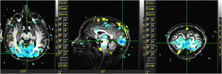
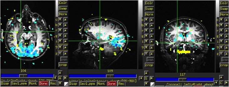
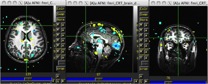
As you can see from the images below there was negative activation in visual cortex during image onset. There was no activation in the amygdala or PFC.
Discussion
Negative activation in visual cortex suggests something is off in either the timing files or contrasts where perhaps we are confusing the ITI with trial onset during either the input of the timing or the contrasts. A lack of activation in the amygdala is not too concerning given this is 1 scan in 1 participant, although a better contrast would be to look at US > fixation. A lack of activation in the PFC is also not concerning given the participants have not been taught to use cognitive restructuring during the first scan. It would be better to analyze data from the second session, as this is when participants are more likely to use cognitive restructuring and their PFC would be engaged.
Conclusions
The only conclusion I can make from my initial attempt at AFNI is that I have a long way to go!! The more I learn the more questions I have, but this has given me a first (rough) pass at pre-processing and data analysis using AFNI. I hope that I will be able to figure out exactly what is going on with my timing and contrasts so I can apply this to future subjects and have better results. I (sort of?) look forward to it!
References
Cox, R (1996). AFNI: Software for analysis and visualization of functional magnetic
resonance neuroimages. Computers and Biomedical Research, 29: 162–173.
Kendall, P.C., & Hedtke, K. (2006). Coping Cat Workbook, 2nd Ardmore, PA: Workbook Publishing.
Klorman, R., Weerts, T. C., Hastings, J. E., Melamed, B. G., & Lang, P. J. (1974). Psychometric description of some specific-fear questionnaires. Behavior Therapy, 5, 401-409.
Schiller, D., Monfils, M.H., Raio, C.M., Johnson, D.C., LeDoux, J.E., & Phelps, E.A. (2010). Preventing the return of fear in humans using reconsolidation update mechanisms. Nature, 463, 49 - 53.
Szymanski, J., & O'Donohue, W. (1995). Fear of spiders questionnaire. Journal of Behavior Therapy and Experimental Psychiatry, 26, 31-34.
Talairach, J., & Tournoux, P. (1988). Co-Planar stereotaxic atlas of the human brain: An approach to medical cerebral imaging, Stuttgart, New York: Thieme Medical Publishers.


