Kaja Johnson
Back to Psych 204 Projects 2009
Foveal and Peripheral Perception in Face and Place Processing
Background
It has been well established in fMRI literature that there are regions in the brain that are selective for particular objects over others, namely faces and places. The Fusiform Face Area (FFA) has been shown to be significantly more active when subjects view faces than other objects (e.g. Kanswisher et al., 1997); the Occipital Face Area (OFA) is known to be responsible for perceiving faces at a basic level (Gauthier et al., 2000), while specifically the rOFA has been implicated in face-part recognition (Pitcher et al., 2007); the Parahippocampal Place Area (PPA) activates more strongly to places than other visual stimuli (e.g. Epstein et al., 1999); the Retrosplenial Cortex (RSc) in involved in spatial learning and memory in animal models (Vann et al., 2003), and has an analogue function in humans in being responsible for processing of the perception of location (Epstein et al., 2007).
The limitation of such studies seeking specialized localization in the brain is that it is still not clear if particular regions of interests (ROIs) are involved in more complex processing rather than this simplistic one-to-one region to function model. Visual stimuli of faces and places may be processed in more comprehensive ways that include taking in information about other factors like location in the visual field. The current study aims to examine this and determine if these specialized regions for face and place processing respond differently to stimuli presented foveally and peripherally. The differential functions of foveal and peripheral vision implicate the possibility that external stimuli may possibly be encoded and processed in different ROIs. Foveal vision is used for examining detailed objects and peripheral vision is used for organizing the broad spatial scene and seeing large objects (Peripheral Vision). Because of the fovea’s responsibility for attention to central details of an expression in the visual field (i.e. eyes, nose and mouth) it is likely that facial processing recruits energy from foveal processing systems. Likewise, because the recognition of a scene or place requires attention to broader context of visual field where details are also maintained in periphery and not solely the fovea, stimuli of this kind should recruit energy from peripheral processing systems. In line with these functions, I hypothesized that 1) face-selective areas (FFA, OFA) will also be selective for foveal stimuli, and 2) place-selective areas (PPA, RSc) will also be selective for peripheral stimuli.
Methods
Subjects
Data was sampled from a session with one subject (female).
MR acquisition
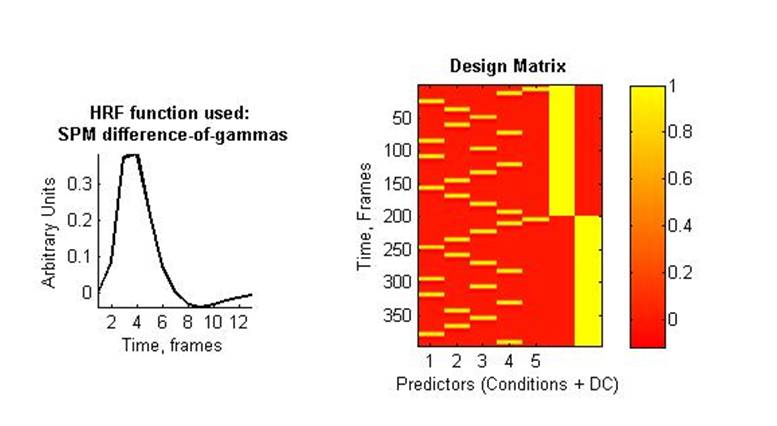
Brain imaging was performed on a 3 tesla whole-body General Electric Signa MRI scanner. During fMRI the subject viewed images of faces of children and adults, abstract objects, cars, indoor scenes, and outdoor scenes presented foveally and peripherally. Stimuli presented in the periphery were larger than stimuli in the fovea to control for size of the object on the retina, as it has been shown that stimuli in the periphery must be larger to be processed at equivalent levels (Latham and Whitaker, 1996). Stimuli were presented in 12 s blocks. Four of each stimulus type were presented (face fovea, face periphery, place fovea, place periphery; 16 total blank blocks were placed between stimulus presentations. Motion and still images were also used to monitor activity in a region known to be not selective for face and place categories (Visual Area Middle Temporal; from here on referred to as MT). The total time of each run was 198 TRs (TR=2s; subjects participated in two 396-s runs with different images.
MR Analysis
The MR data was analyzed using mrVista software tools.
Pre-processing
All data were slice-time corrected, motion corrected, and repeated scans were averaged together to create a single average scan for the subject.
Model
I estimated a GLM with two contrasts to generate parameter maps for analysis: one for the face-selective areas and the other for place-selective areas. For face-selective areas this was child and man as active condition and indoor, outdoor, cars and abstract object as the control condition. For place-selective areas this was indoor and outdoor as active condition and child, man, cars, and abstract object as the control condition. For the control scans in the MT the contrast was motion as the active condition and still images as the control condition.
Results
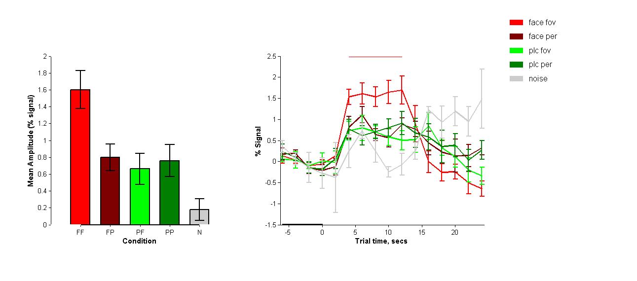
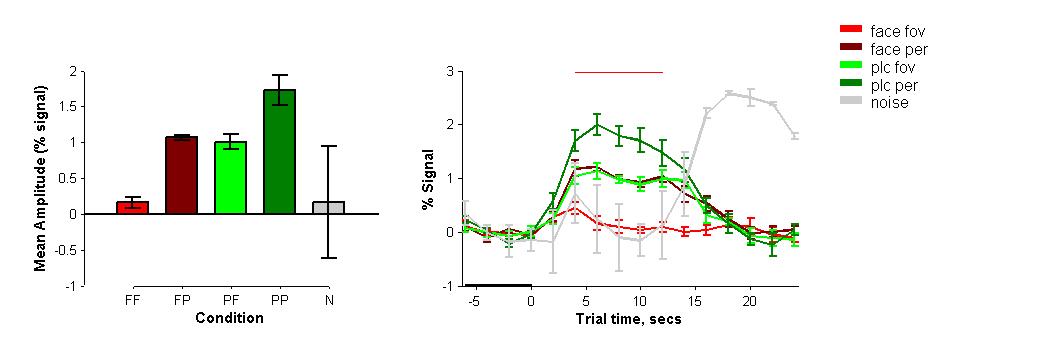
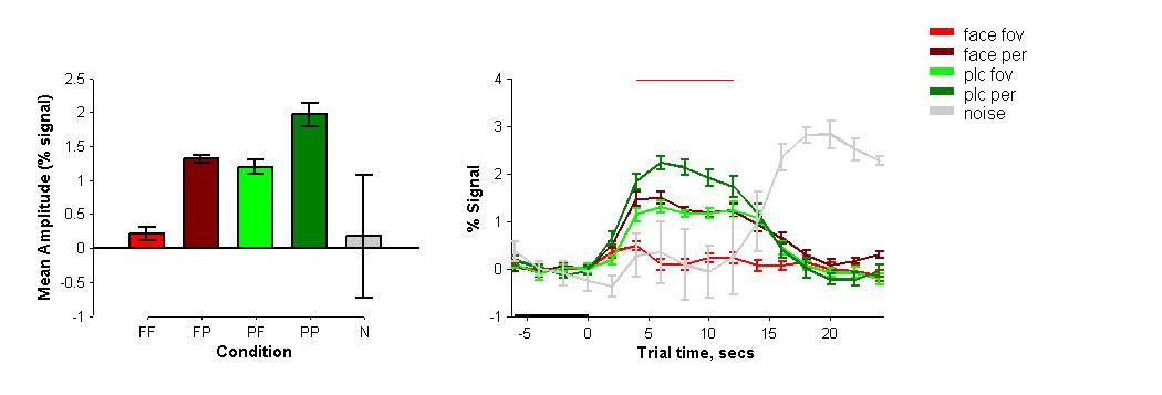
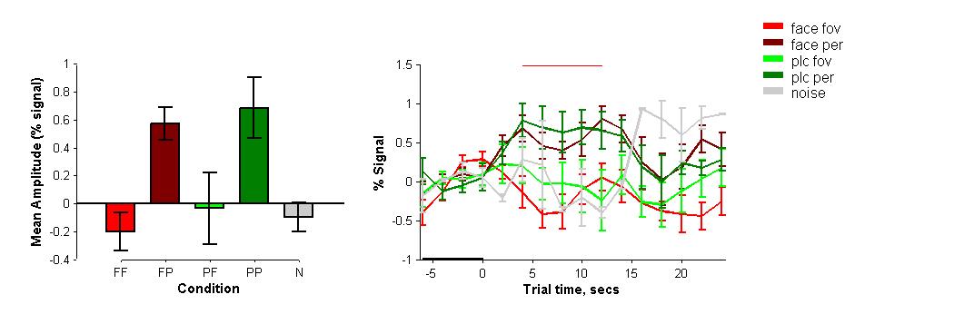
As expected the foveal presentations of face and place stimuli in all ROIs follow findings of previous research as stimuli in studies are traditionally only presented in the fovea. Face-selective areas showed greater activity when presented with faces than with places, and the reverse is true for place-selective areas.
FFA
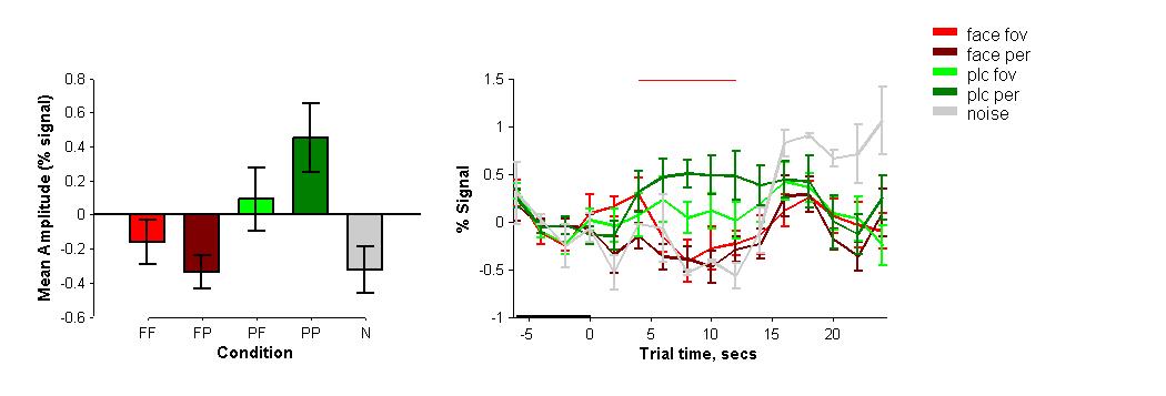
In the left hemisphere of the brain, the FFA appears to be particularly selective for foveally presented stimuli of faces, and processes peripheral presentations of faces similarly to that of place stimuli. This implies that the left FFA is more specifically selective for faces presented in particular areas of the visual field, and not faces indiscriminately. The right FFA did not reveal any differences across all stimuli and did not show any selectivity for places, however it is often the case that this selectivity is localized to one hemisphere.
OFA
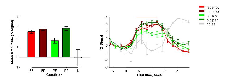
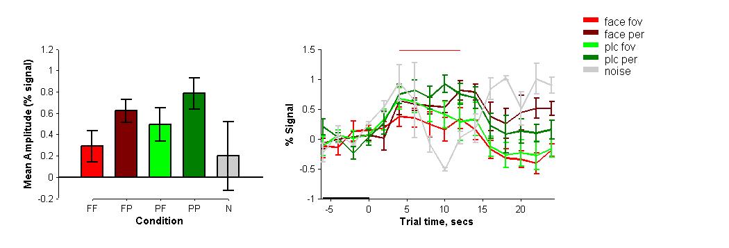
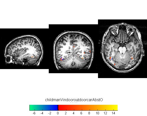
The left and right OFAs show selectivity for faces, however it appears the lOFA is particularly selective for foveal stimuli in general, including places (there was more activation for foveal places than peripheral places), while the rOFA is particularly selective for peripheral stimuli (again, a main effect of location was seen with more activation for both faces and places in the periphery).
PPA
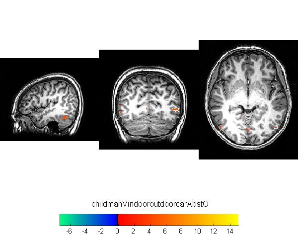
The left and right PPAs showed selectivity for peripherally presented stimuli across categories. Again, it maintained expected selectivity for places controlling for location of the presentation.
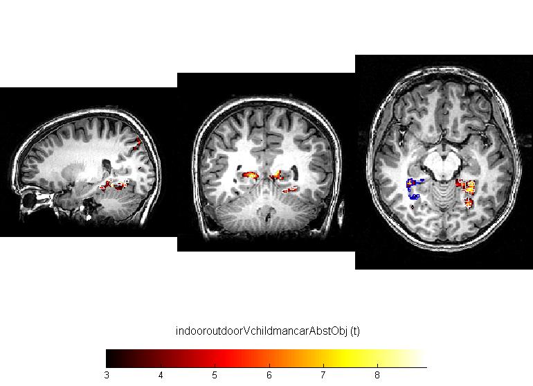
RSc
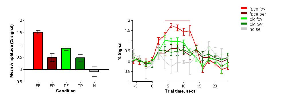
The left and rigth RSc regions do not appear to differentiate between foveal and periphery stimuli as the activity across locations showed only a main effect of category. However, this face-place distinction seems to occur in the periphery, as the activations were driven by the results seen when stimuli for faces and places were presented in peripheral locations.
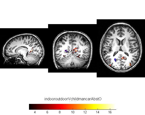
MT
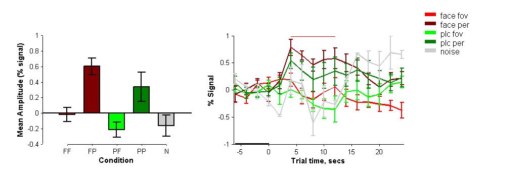
This served as an effective control condition. There were so particular significant selectivities for categories since this region responds to motion stimuli. However, the MT also seems to prefer peripheral stimuli over foveal ones as there were greater activations during these presentations.

Conclusions
Localization for specialized processing generally follows expectations based on functionality in the external world. For example, it makes sense for the MT to be selective for peripheral stimuli and motion stimuli, because it will be most adaptive to quickly respond to potentially dangerous objects coming rapidly from surprise areas to your sides (e.g. an oncoming lion or fist). Likewise, face-selective areas specialize for foveal processing due to the need to focus attention on central details of the visual field. Place-selective areas specialize for peripheral processing due to the need to focus on details located on all parts of the visual field for accurate recognition. Results in the right hemisphere across categories of faces and places seemed to prefer peripheral processing which contradicts the original hypotheses. This, however, makes sense as the right side of the brain is also known to specialize in spatial processing, so movement and place are important areas of focus for activities that require spatial skills. The findings also implicate the RSc as a possible area where general processing of peripheral visual stimuli occurs, which may contribute specificity to the function of this region.
The limitation of this study is of course the small number of subjects examined. In order to generalize these results, replication must be done in a larger pool of participants.
References
Epstein, R., Harris, A., Stanley, D. and Kanwisher, N., 1999. The parahippocampal place area: recognition, navigation, or encoding?. Neuron 23, pp. 115–125.
Epstein RA, Parker WE, Feiler AM (2007) Where am I now? Distinct roles for parahippocampal and retrosplenial cortices in place recognition. J Neurosci 27:6141–6149.
Gauthier I, Trends Cognit. Sci. 4, 1 (2000)
Kanwisher N, McDermott J, Chun MM (1997 Jun 1). "The fusiform face area: a module in human extrastriate cortex specialized for face perception". J Neurosci. 17 (11): 4302–11.
Latham K., Whitaker D. A comparison of word recognition and reading performance in foveal and peripheral vision (1996) Vision Research, 36 (17), pp. 2665-2674.
Peripheral Vision. (n.d.). WebExhibits. Retrieved March 16, 2010, from http://www.webexhibits.org/colorart/ag.html
Pitcher D, Walsh V, Yovel G, Duchaine B. TMS evidence for the involvement of the right occipital face area in early face processing. Curr Biol doi:10.1016/j.cub.2007.07.063, 2007.
Vann S.D., Wilton L.A.K., Muir J.L., Aggleton J.P. Testing the importance of the caudal retrosplenial cortex for spatial memory in rats (2003) Behavioural Brain Research, 140 (1-2), pp. 107-118.
